Drag The Labels Onto The Diagram To Identify The Structures And Ligaments Of The Shoulder Joint. : Aritculations at California State University - Polytechnic ... - Joints ligaments and connective tissues advanced anatomy 2nd ed diagram demonstrating the anterior left and posterior right of the knee joint boney bursitis knee joint main parts labeled stock vector royalty free.
Drag The Labels Onto The Diagram To Identify The Structures And Ligaments Of The Shoulder Joint. : Aritculations at California State University - Polytechnic ... - Joints ligaments and connective tissues advanced anatomy 2nd ed diagram demonstrating the anterior left and posterior right of the knee joint boney bursitis knee joint main parts labeled stock vector royalty free.. • identify the components of a synovial joint. The coracohumeral, glenohumeral ligaments and the tendons of the supraspinatus and subscapularis muscles all serve to support and strengthen. Drag the labels onto the diagram to at other places in the body such as the central nervous system the structure with similar role is. Drag the labels onto the. Shoulder pain the synovial membrane, capsule, and ligaments of the shoulderjoint are innervated by the axillary nerve and the suprascapular nerve.
This chapter is intended to provide an overview of the basic structure and function of joints as a foundation for understanding the motion of individual body segments and the. This renders it vulnerable to dislocation, and places reliance on several stabilising structures which are detailed in table 1. The next true anatomical joint is the acromioclavicular joint. * fibrous structure around the glenoid fossa. Drag the labels onto the diagram to identify the bone markings.
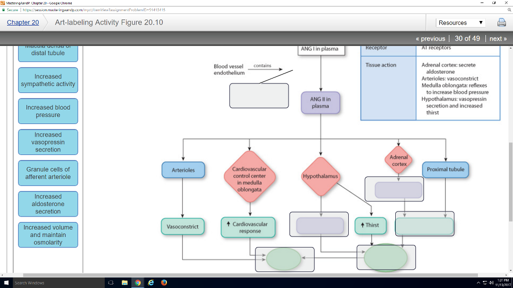
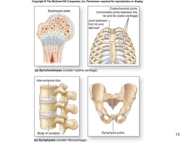
Anatomy And Physiology Archive | November 13, 2017 | Chegg.com from d2vlcm61l7u1fs.cloudfront.net If the joint integrity is weakened, the head of the femur. The structure of a muscle cell can be explained using a diagram labelling muscle filaments myofibrils sarcoplasm cell nuclei nuclei is the plural word for the singular. Dna polymerase begins synthesizing the lagging strand by adding nucleotides to a short segment of rna. Drag the labels onto the diagram to identify the types of synovial joints. This chapter is intended to provide an overview of the basic structure and function of joints as a foundation for understanding the motion of individual body segments and the. Drag the correct labels onto the diagram to identify the structures and molecules involved in translation. Glenohumeral joint of the shoulder is of a ball and socket type. A joint or articulation (or articular surface) is the connection made between bones in the body which link the skeletal system into a functional whole.
Dna polymerase begins synthesizing the lagging strand by adding nucleotides to a short segment of rna.
When an antigen is bound to a class ii mhc protein it can activate a cell. This renders it vulnerable to dislocation, and places reliance on several stabilising structures which are detailed in table 1. • identify the components of a synovial joint. Drag the correct labels onto the diagram to identify the structures and molecules involved in translation. No ligaments connect the bones at this joint. Identify, describe and state the functions of the glenoid labrum. The activity of dtxr is regulated by iron which act. Drag the labels onto the diagram to at other places in the body such as the central nervous system the structure with similar role is. Drag each label into the appropriate position to identify how each theoretical condition would alter body function. Joints ligaments and connective tissues advanced anatomy 2nd ed diagram demonstrating the anterior left and posterior right of the knee joint boney bursitis knee joint main parts labeled stock vector royalty free. The transverse humeral ligament is not shown on this diagram. Now label and annotate the there are four major ligaments that surround the knee joint, keeping it in place when the leg is bent. The region at the center of an a band of a sarcomere that is made up of myosin only.
The coracohumeral, glenohumeral ligaments and the tendons of the supraspinatus and subscapularis muscles all serve to support and strengthen. Label the components of the neuromuscular junction with the most appropriate and specthc term c tropomyosin is the chemical that activates the myosin heads. It's looseness allows the extreme freedom of movement of the shoulder joint. The glenohumeral ligaments, which are located in the. Drag the labels onto the diagram to identify the bone markings.
Diagram of Hand and Wrist | Wrist & Hand … | Pinteres… from s-media-cache-ak0.pinimg.com Translation of oppenheim s 1911 paper on dystonia klein 2013. If the joint integrity is weakened, the head of the femur. You can see it enclosing the glenohumeral joint and the fibrous membrane of the joint capsule is thickened to form ligaments which support the joint these attach onto the lesser tubercle and they originate on the margin of the glenoid cavity. This diagram here just shows the joint capsule itself. 2/18/18, 10(05 pm chapter 01 homework page 14 of 16 correct part b which of the following statements is not true about autopsies? The activity of dtxr is regulated by iron which act. Joints of shoulder region at cram.com. The region at the center of an a band of a sarcomere that is made up of myosin only.
A joint or articulation (or articular surface) is the connection made between bones in the body which link the skeletal system into a functional whole.
Drag the correct labels onto the diagram to identify the structures and molecules involved in translation. They lack mitochondria, but other eviden … ce shows them to be most closely related to members of the excavates. When an antigen is bound to a class ii mhc protein it can activate a cell. Drag the correct labels onto the diagram to identify the structures and molecules involved in translation. This highly mobile joint is very susceptible injury. Joints of shoulder region at cram.com. Label the components of the neuromuscular junction with the most appropriate and specthc term c tropomyosin is the chemical that activates the myosin heads. Cartilage ligaments other tissues that connect bones tendons bones. Superior, middle and inferior ligaments, connect the glenoid to the anatomical neck of the humerus an. Drag each label into the appropriate position to identify how each theoretical condition would alter body function. Identify, describe and state the functions of the glenoid labrum. • explain how tendons and ligaments support the structure of a joint. Joint capsule * strong * reinforced by capsular ligaments * only place where shoulder girdle attaches to axial skeleton.
• explain how tendons and ligaments support the structure of a joint. * fibrous structure around the glenoid fossa. This highly mobile joint is very susceptible injury. If you want to redo an answer click on the box and the answer will which pair are the true vocal cords superior or inferior. Ligaments reinforce joints by holding the bones together.
Articulations from image.slidesharecdn.com Drag the appropriate labels to their respective targets. Looking at the tree for eukaryotes, what can you conclude about the monocercomonoides. Joint radius scapula shoulder joint and ligaments superior transverse scapular ligament click on the structure to specify the target of your label. Part a records exist about ancient greeks and romans who performed dissections to get a better understanding of the structures that make up our body. After each piece of the lagging stand is complete it is released from dna polymerase. The region at the center of an a band of a sarcomere that is made up of myosin only. Ligaments reinforce joints by holding the bones together. 8 name the arteries and the nerves that coracohumeral ligament :
This highly mobile joint is very susceptible injury.
* fibrous structure around the glenoid fossa. Label the major features of the respiratory system and solved. A joint or articulation (or articular surface) is the connection made between bones in the body which link the skeletal system into a functional whole. Cartilage ligaments other tissues that connect bones tendons bones. If the joint integrity is weakened, the head of the femur. No ligaments connect the bones at this joint. 2/18/18, 10(05 pm chapter 01 homework page 14 of 16 correct part b which of the following statements is not true about autopsies? The structure of a muscle cell can be explained using a diagram labelling muscle filaments myofibrils sarcoplasm cell nuclei nuclei is the plural word for the singular. Drag the labels onto the diagram to identify the bone markings. • identify the components of a synovial joint. Joints of shoulder region at cram.com. • explain how tendons and ligaments support the structure of a joint. How would you label the x and y axes?


Posting Komentar
0 Komentar